CHIROPRACTIC
Chiropractic X-ray Software – XRAY Solutions delivers the most advanced and complete chiropractic digital imaging viewing solutions with its DR flat panel detectors and ChiroView software. With our extensive flat panel detector manufacturing knowledge, Rayence is able to offer a custom, retrofit DR configuration without modifying your existing equipment making installation quick and economical.
This cost-effective design allows a facility to maximize its investment. Our 17×17 inch detector gives you a full field of view not available with X-ray film or CR.

Products
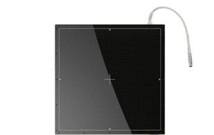
Tethered DR Flat Panel Detector
Tethered DR Flat Panel Detector 1717SCC is an economical solution that allows the user to experience the benefits of flat panel Digital Radiography.
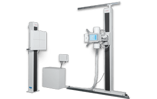
Complete Digital XRAY Room
One of the most affordable
upright systems that incorporates safety, reliability, value, quality and ease of use. System includes features that will increase patient thru-put and minimize the time needed for exams.
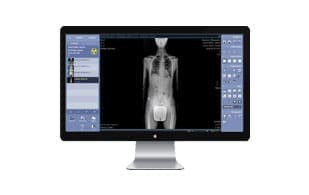
Acquisition Software
Designed to provide user-friendly functionality and increase workflow, ChiroView allows image viewing to be done. Includes full spine stitching and measurement tools with in software guide
1717SCC/SGC • 17×17 Tethered Flat Panel Detector
UNIVERSAL AND AFFORDABLE SOLUTION WITH LARGE COVERAGEN
The detector possesses the thickness of traditional ISO 4090 film cassettes, which makes retrofitting them into standard cassette trays easy. The use of 17×17 inch detector provides more flexibility when positioning anatomy and eliminates the need to be rotated for certain.
The 1717 S-Series is constructed with Rayence’s auto-trigger signal sensing technology, removing the need for generator integration. Efficiency is improved by streamlining workflow through the elimination of the additional steps required when using film or CR.
Together with an image preview time of less than 5 seconds, patient throughput and overall productivity is increased, and the wait time for patients is decreased, providing an economical and ideal solution for any X-ray room.

Features
| Auto-Trigger Signal Sensing Technology |
| 17×17 inch Active Area |
| Ideal for Table and Upright Wall Stands |
| Lightweight and Thin Dimensions |
| 127um Pixel Pitch |
Features
| Detection Area | 17 x 17 inch |
| Sensor Type | Amorphous Silicon with TFT |
| Scintillator | Cesium (1717SCC) / Gadox (1717SGC) |
| Pixel Matrix | 3328 x 3328 |
| Pixel Pitch | 127 um |
| A/D Conversion | 14/16 bit |
| Resolution | Max. 3.9lp/mm |
| Preview Time | ≤ 2Sec. |
| Energy Range | 40~150kVp |
| Data Output | Ethernet 1Gbps |
| Weight | 9.25lbs / 4.2kg |
| Dimensions | 18.1 x 18.1 x 0.6in |
Film/Digital Imaging Solutions • CH1 17” X 17” UPRIGHT IMAGING
Our fixed 40” or 72” SID upright system further reduces the cost of ownership by eliminating the floor and wall tracks supplied on a traditional system. This configuration is ideal for any practice that has limited space and can accomplish their imaging requirements at a fixed 40” or 72” SID. Magnetic locks for vertical movement of the tube and receptor insures ease of positioning for you and your patient.
Features
| 13-SM/S105C Floor Wall Mount Tube Stand |
| Platform tube mount, 90◦ tube rotation |
| Magnetic locks control vertical movement (mechanical lock for tube rotation) |
| 10” (25.4 cm) Arm minimizes room size requirement |
| Angle indicator |
| 60” (152.4 cm) Floor Track |
| 13-BF7-GC/S109 Wall Stand |
| Floor wall mounted |
| 9.75” to 72.75” (24.8 to 184.8 cm) Vertical travel |
| 10” (25.4 cm) Arm minimizes room size requirement |
| Magnetic locks |
| 17” x 17” Grid cabinet |
| 103 line, 10:1 ratio grid |
| Cassette tray |
| E7239FX X-Ray Tube |
| 140,000 Heat units |
| 1.0 x 2.0 mm focal spots |
| Other tubes optional |
| CML125-0001-C Collimator |
| Certified manual collimator |
| 30 to 120 second adjustable light on timer |
| Laser light cross hairs |
| LED light source |
| Retractable tape measure |
| Swivel mount |
| 10539-20 High Tension Cables |
| 20’ (609.6 cm) Long |
Features
| 14”x17” (35 x 43 cm) or 17”x17” (43 x 43 cm) Digital panel |
| Various grids |
| Tilt mechanism |
| 14” (35.5 cm) or 23”(58.4 cm) Tube arm |
| 96” or 120” (243.8 or 304.8 cm) Floor track |
| Fixed SID |
Acquisition Software
It is designed to provide user-friendly functionality and increase workflow. Xmaru ChiroView allows image viewing to be done in every exam room throughout the office.
The software also allows users to import DICOM images from outside facilities, burn images to a CD/DVD or USB memory stick, and print to paper. With our industry leading chiropractic measurement tools and easy to follow tutorials on how to properly apply the measurement overlays, Rayence provides Chiropractors a sensible and economical Digital X-ray solution.

Features
| This stand-alone viewer comes with the same features as the XmaruView, plus 55 more |
| The Xmaru ChiroViewer comes with standard features like, DICOM Send, Image Stitching, DICOM Print, Export/Import JPEG and DVD/CD Burning with viewer |
| For disaster recovery, the Xmaru ChiroViewer is built with features like image backup/restore functions |
| An important feature on the Xmaru ChiroViewer is the short tutorial for measurment tools: allowing users not familiar with how to “draw” the measurement to become complete professionals |
| Each tutorial explains the function and usage of the tool. In addition, you can edit and make your own description for your tools |
Features
| Skull |
| Vastine-Kinney Method of Pineal Gland Localization |
| Sella Turcica Size |
| Basilar Angle |
| McGregor’s Line |
| Chamberlain’s Line |
| Macrae’s Line |
| Height Index of Klaus |
| Boogard’s Line |
| Boogard’s Angle |
| Anterior Atlanto-Occipital Dislocation Measuremen |
| Lumbar Spine |
| Intervertebral Disc Height (Hurxthal’s Method) |
| Intervertebral Disc Height (Farfan’s Method) |
| Lumbar Intervertebral Disc Angles Lumbar Lordosis |
| Lumbosacral Lordosis Angle |
| Sacral Inclination |
| Lumbosacral Angle |
| Lumbosacral Disc Angle |
| Lumbar Gravity Line |
| Lateral-Bending Sign |
| Meyerding’s Grading Method in Spondylolisthesis |
| Ullmann’s Line |
| Eisenstein’s Method for Sagittal Canal Measurement |
| Intercrestal Lineo |
| Cervical Spine |
| Atlantodental Interspace(ADI) |
| Method of Bull George’s Line |
| Posterior Cervical Line |
| Sagittal Dimension of the Cervical Spine Canel |
| Cervical Gravity Line |
| Cervical Lordosis (Angle of the Cervcal Curve) |
| Cervical Lordosis (Depth Measurement) |
| Cervical Lordosis (Method of Jochumsen) |
| Stress Lines of the Cervical Spine(Flexion) |
| Stress Lines of the Cervical Spine(Flexion) |
| Prevertebral Soft Tissueso |
| Thoracic Spine |
| Cobb’s Method of Scoliosis Evaluation |
| Risser-Ferguson Method of Scoliosis Evaluation |
| Thoracic Kyphosis |
| Thoracic Cage Dimension |
| Lower Extremity |
| Teardrop Distance |
| Hip Joint Space Width |
| Acetabular Depth |
| Center-Edge Angle |
| Symphysis Pucis Width |
| Presacral Space |
| Acetabularr Angle |
| Iliac Angle and Index |
| Measurements of Protrusio Acetabuli |
| Femoral Angle |
| Skinner’s Line |
| Axial Relationships of Knee |
| Upper Extremity |
| Glenohumeral Joint Space |
| Acromiohumeral Joint Space |
| Acromioclavicular Joint Space |
| Special |
| Gonstead Measurements(Pelvis) |
| Cervical Curve |